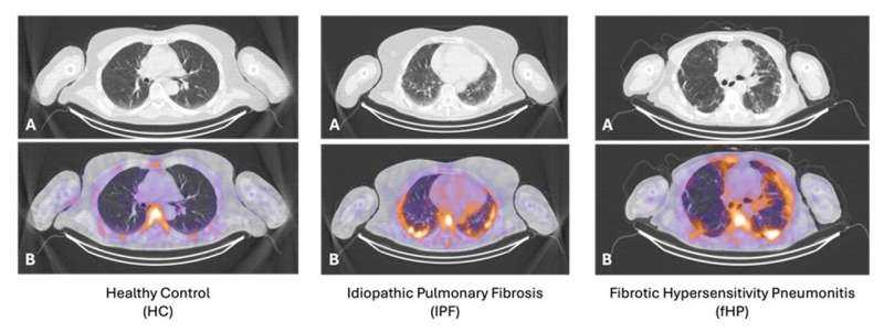
A groundbreaking SPECT/CT imaging approach can now accurately differentiate inflammation from fibrosis in interstitial lung disease patients, aiding in targeted treatment decisions.
A new SPECT/CT (Single Photon Emission Computed Tomography/Computed Tomography) imaging technique has shown promising results in accurately distinguishing between inflammation and fibrosis in interstitial lung disease (ILD) patients. This advancement was presented at the Society of Nuclear Medicine and Molecular Imaging 2026 Annual Meeting, held from May 30 to June 2 in Los Angeles.
The research highlights that this molecular imaging approach can provide critical information for healthcare providers to determine which ILD patients would benefit most from anti-inflammatory treatments. By identifying inflammation specifically, doctors can tailor treatment plans to target the underlying causes of lung disease, potentially reducing unnecessary side effects associated with inappropriate therapies.
This new technique could significantly improve patient outcomes by ensuring that those who need anti-inflammatory medications receive them while minimizing exposure to drugs that may only exacerbate fibrotic conditions. The ability to accurately differentiate between these two key components of ILD is crucial for optimizing treatment strategies and improving the quality of care for patients suffering from this complex condition.
The Society of Nuclear Medicine and Molecular Imaging 2026 Annual Meeting brought together experts in nuclear medicine and molecular imaging, providing a platform for sharing cutting-edge research like this SPECT/CT approach. This development represents an important step forward in personalized medicine for ILD, offering hope for more precise and effective treatment options in the future.