University Medical Center Groningen has developed a new method to produce a crucial PET scan tracer more efficiently. This innovation allows for significantly larger numbers of patients to be examined, shortening wait times and enabling earlier diagnosis.
University Medical Center Groningen (UMCG) has introduced a groundbreaking new production process that simplifies the creation of a specific radioactive tracer used in Positron-Emission Tomography (PET) scans. This advancement is particularly significant for diagnosing conditions such as Parkinson's disease and neuroendocrine tumors, where quick and accurate diagnoses are crucial.
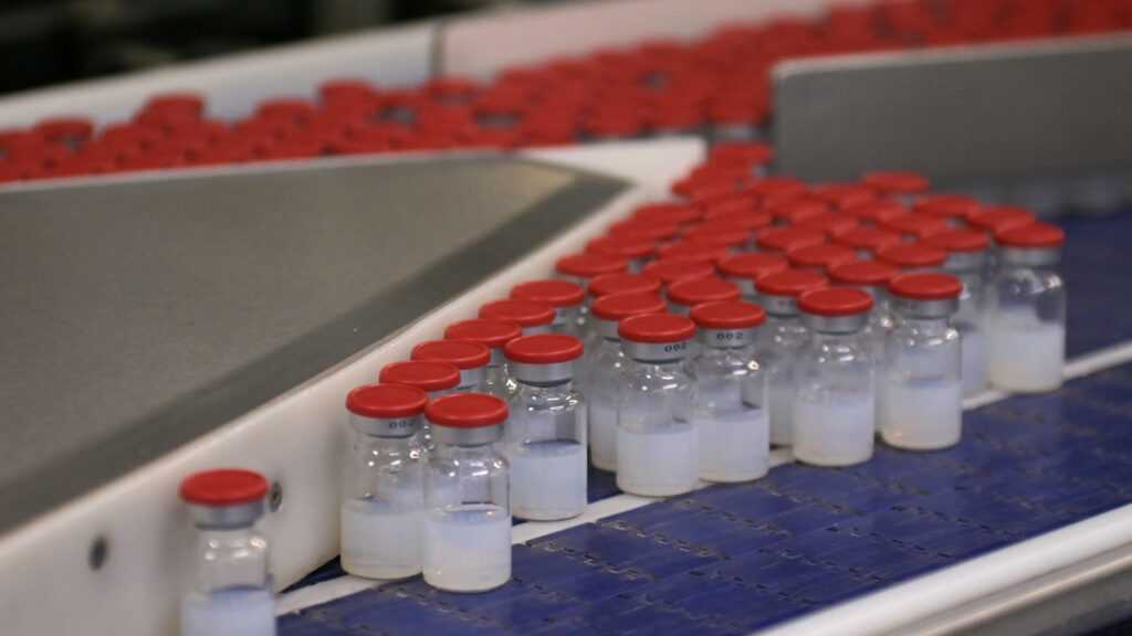
The PET scan tracers, including [18F]fluorodopa (FDOPA), play a vital role in visualizing and measuring biological processes within the body. By injecting a small amount of this tracer into patients, doctors can assess various functions like energy consumption or metabolic activity, which is particularly useful for detecting tumors or evaluating brain function.
Traditionally, producing FDOPA was a complex and time-consuming process that limited its availability to only a few patients at a time. However, the UMCG's new method has streamlined this procedure significantly. The production now involves fewer steps and uses a liquid form of radioactive gas instead of gas, making it both safer for staff and more efficient.
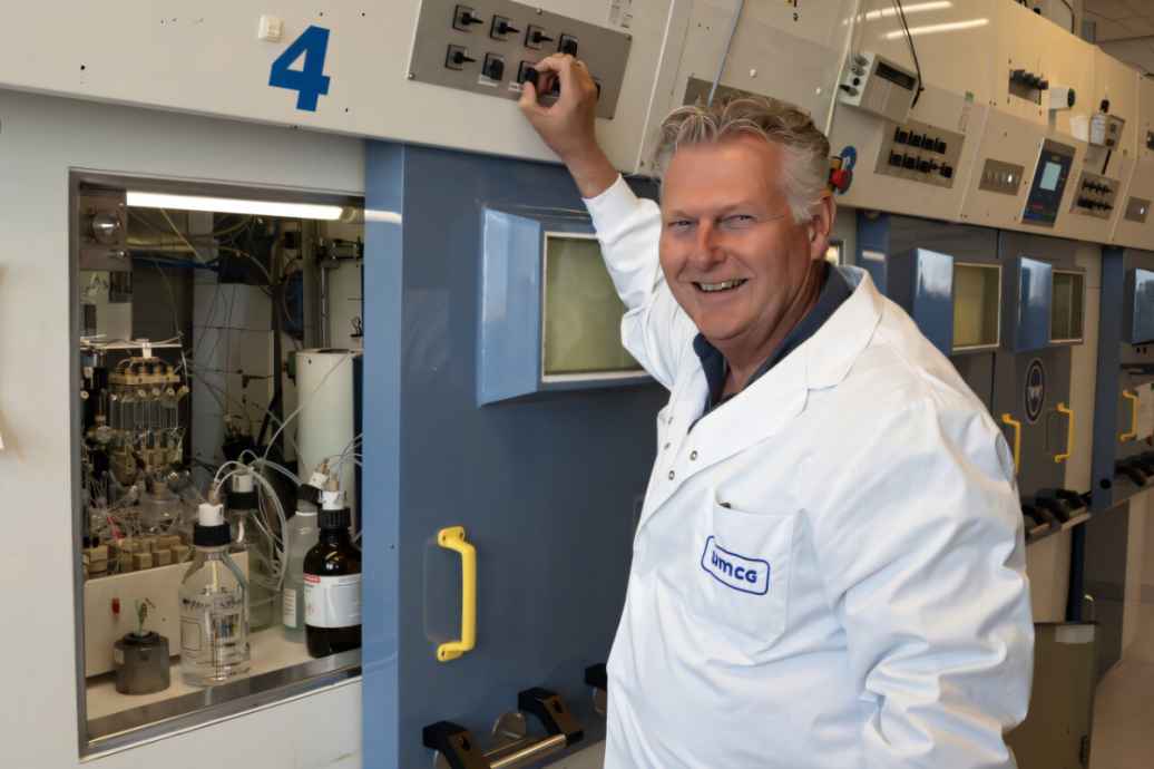
Gert Luurtsema, a clinical radiochemist from the Department of Nuclear Medicine and Molecular Imaging at UMCG, explains that this innovation not only reduces the number of required steps but also makes the process much quicker. "Previously, we could help five to six patients per batch; now, with our new method, we can produce up to twenty to thirty doses in a single production run," he notes.
This increase in production capacity is expected to have a substantial impact on patient care. Dr. Luurtsema anticipates that the shorter wait times will enable earlier diagnoses and quicker treatment initiation for conditions like Parkinson's disease and cancer. "By shortening waiting periods, we can provide patients with clarity sooner and start them on treatment faster," he adds.
The development of this new production method took over ten years, during which UMCG leveraged its extensive expertise in radiochemistry along with collaborations from other research groups, including Nobel laureate Ben Feringa's team. This interdisciplinary approach ensured that the tracers were not only developed but also rapidly tested and implemented in clinical settings.
The UMCG was one of the first centers in the Netherlands to have a medical cyclotron—a device used to produce radioactive isotopes essential for PET scans. These isotopes form the basis for the tracers needed in diagnostic imaging, and their short half-life necessitates close proximity between production facilities and patients.
Last month marked the first time FDOPA tracer was produced using this new method and administered to patients. Dr. Luurtsema is optimistic about the potential benefits of this innovation. "This will significantly improve patient care by reducing wait times and enabling quicker diagnoses," he says. "For conditions like Parkinson's disease, where early intervention can make a substantial difference in quality of life and prognosis, this development could be transformative."
The new method has been patented, marking another milestone for UMCG’s ongoing commitment to advancing medical diagnostics and treatment options. As the global population ages, the demand for PET scans is expected to grow, making this innovation even more valuable.
In conclusion, University Medical Center Groningen's breakthrough in producing FDOPA tracers more efficiently represents a significant leap forward in patient care. This new capability promises not only faster diagnoses but also enhanced treatment outcomes and improved quality of life for patients suffering from brain diseases and other conditions requiring PET scans.